

The Concept of the Disease
Classical Hodgkin Lymphoma (CHL) is a tumour of activated B-cells that have lost most of the immunophenotypic characteristics of their normal counterparts. A defining characteristic of CHL is the tumour cells often constitute a minority of the volume of the tissue mass, which consists mainly of normal haemopoietic cells and fibrous tissue.
The term classical is used to make a distinction from nodular lymphocyte predominant Hodgkin Lymphoma which is biologically and clinically distinct.
How is it Diagnosed?
Lymph Node Biopsy
- The normal structure of the node is replaced by tumour.
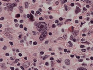
- At least a proportion of the tumour cells have highly characteristic morphology- Reed Sternberg cells
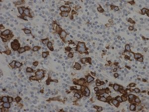
- The tumour cells express activation markers (CD30 and IRF4) but the normal B-cell lineage markers (CD20, CD19 or CD79) are usually absent.
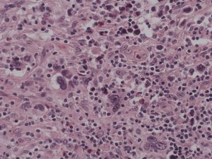
- The tumour cells, which comprise the minority, are associated with large numbers of normal macrophages, lymphocytes and eosinophils and in most cases there will be varying degrees of fibrosis.
Lymph Node Morphology – Reed Sternberg cells
Immunocytochemistry
Pattern of Disease
- The distribution of disease is highly characteristic in CHL and most patients have disease centres on the mediastinum with contiguous spread to other lymph node groups.
- Involvement of tissues other than lymph nodes is rare.
What is the Clinical Outcome?
About 75% of patients treated with standard chemotherapy, and radiotherapy where indicated, are cured of their disease and survive for more than 5 years. Many of those who relapse can still be effectively treated.
Age is an important prognostic factor and data on the outcome of older patients is limited. The Hasenclever index is the standard method of assessing prognosis at presentation.
| Score | Hasenclever Risk Group |
| ≤2 | Low |
| ≥3 | Moderate to High |
| Risk Factor | Score +0 | Score +1 |
| Serum Albumin | ≥ 40 g/L | > 40 g/L |
| Haemoglobin | ≥ 10.5 g/dL | < 10.5 g/dL9/L |
| Age | <45 | ≥ 45 |
| Stage (Ann Arbor) | I to III | >IV |
| WBC | < 15 x 109/L | ≥ 15 x 109/L |
| Lymphocytes | ≥ x 109/L ≥ 8% of differential |
< 0.6 x 10⁹/L < 8% of differential |
| Sex | Female | Male |
PET scans are used to assess response to chemotherapy and using this data to modify subsequent treatment is being evaluated in clinical trials.