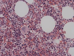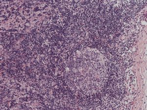

Microscopic examination of tissue sections plays an important role in the diagnosis of haematological malignancy. Bone marrow and lymph nodes have a complex internal structure which reflects the way that cells interact with each other during normal haemopoiesis ( the continuous production of new peripheral blood cells in the bone marrow) or during the response of immune system to foreign material (the main function of lymph nodes).
Changes in the interaction between malignant and normal cells are a fundamental property of all types of haematological malignancy. This can be observed microscopically by identifying changes in the relative proportions of different types of cell, the breakdown of the internal structure of tissue and the detection of cells in locations where they would not normally be found.
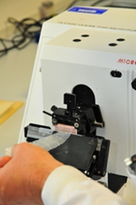
Preparing a Biopsy for Histological Examination
 Microscopic examination of tissue requires the preparation of very thin translucent sections. For soft tissues such as lymph node these would be typically cut at 4 microns and for bone marrow 1-2 microns. These sections are stained to dyes or using antibodies (immunocytochemistry) that differentially highlight individual components of cells or tissues.
Cutting sections with this degree of precision requires the specimen is first processed to produce a block with uniform consistency and hardness. This is carried out in two stages.
- The tissue is chemically fixed with formalin to preserve the internal structure
- All of the water is removed from the specimen and replaced by paraffin wax for soft tissues or methyl methacrylate resin for bone marrow. The presence of bone in bone marrow biopsies means that a much harder material must be used.
It is important to realise that typically this process takes 48 hours to complete and this is a major component on the time taken to report the specimen.
Staining the Sections
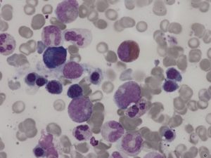
The diagnosis of haematological malignancies relies heavily on the identification of patterns of protein expression by the cells under examination. Immunocytochemistry uses antibodies labelled coloured dyes are used as specific probes to localise specific protein molecules to individual cells in tissue sections. This is used both to accurately individual cell types in tissue and to look for the presence of abnormal patterns of protein expression that distinguish malignant from normal cells. Immunocytochemistry is complementary to flow cytometry.
Examples